![]()
Prevention is better than Treatment
R&D
DIAGNOSTICS LTD®
SQMMR500
& SQMMR1250
INSTRUCTIONS FOR USE (PACKAGE INSERT)
A
SCREENING KIT FOR DRIED BLOOD SPOTS & WHOLE BLOOD SAMPLES
This kit is particularly suitable for
screening for G-6-pd deficiency
in newborns.
KIT SIZE: 10 x 50
TESTS & 5 x 250 TESTS
Manufactured
by :
R&D
DIAGNOSTICS LTD.
41, El. Venizelou str.,
15561 Holargos
Greece
www.rddiagnostics.com
Catalog No : SQMMR500 & SQMMR1250
Procedure
The
assay procedure is according to reaction described by Beutler (1,2).
The enzyme determined is glucose-6-phosphate dehydrogenase which is
abbreviated either as G-6-PD or G-6-PDH.
Test
principle
G-6-PD
Glucose-6-P
+ NADP+
------->
gluconate-6-P
+ NADPH + H+
The
NADPH produced in the reaction fluoresces under long-wave UV-light.
If there is a marked deficiency of this enzyme, or if G-6-PDH is
lacking entirely, no fluorescence will be observed.
Sample
material
See
noted # 1 and 2.
Whole blood dried on filter paper (3).
Apply a drop of blood to absorbent paper ("Gurthie Test Paper", Schleicher & Schuell No. 2992) and let dry completely (atable for one week at 20-25oC).
Whole blood.
Whole blood may be used instead of dried blood. Heparin, citrate, oxalate and EDTA are suitable anticoagulants. The blood specimens are stable for seven days at most. Use 0,005 ml (5 microliters) for the assay.
Reagents
Contents
of solution Concentration in the test
Glucose-6-phosphate
1 mmol/l
NADP
0,75 mmol/l
GSSG
(oxidized glutathione) 0,8 mmol/l
Saponin
0,2%
Tris(hydroxymethyl)-
aminomethane
225 mmol/l, pH 7.8
Preparation
and stability of reagent solution
Dissolve
the contents of the vial containing the freeze dried powder (Reagent
vial; code RD7002) with 5 ml from a Dilution buffer vial (code
RD7001). Please note that the Dilution buffer vial contains more
than 5 ml buffer. Please also note that the Reagent vial has been
sealed under vacuum to allow for a better stability.
Stable
for four weeks at +4oC
two months at -20oC
Sample
preparation
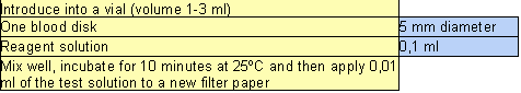
Punch
out a disk of blood-stained paper of 5 mm diameter (3 mm can also be
used).
Procedure (see chart)
Evaluation
When
the filter paper is dry (approximately after 1 hour), view under a
long-wave UV-lamp in a darkened room (see note # 3). Samples
obtained from normal or slightly reduced G-6-PDH activity will show
strong fluorescence. Failure to fluoresce after a 10 minute
incubation suggest a total or marked deficiency of G-6-PDH.
Please note
In some forms of G-6-PDH deficiency, young erythrocytes manifest normal enzyme activity. Blood from patients who have just experienced a hemolytic crisis must first be treated by the procedure described by Herz et al (4) to separate the older erythrocytes from the prevailing population of young ones. Use 0,005 ml of the suspension so obtained for the assay.
If the patient has received a blood transfusion, this test is clinically significant only after 30 days have elapsed, because the donor's erythrocytes generally manifest a normal G-6-PDH activity and can thus bias the result before the expiration of this time.
Any commercially available UV-lamp emitting long-wave UV-light is adequate for the evaluation.
Warning: Dilution buffer contains sodium azide as preservative. Do not swallow. Avoid contact with the skin and mucous membranes.
References
Beutler E. Drug-induced hemolytic anemia and non-spherocytic hemolytic anemia. In Glucose-6-Phosphate Dehydrogenase (Yoshida A. and Beutler E., Eds) pp. 3-12, Academic, Orlando.
Beutler E. A series of new screening procedures for pyruvate kinase deficiency, glucose - 6 - phosphate dehydrogenase deficiency and glutathione reductase deficiency. Blood 1966;28:553-562.
Dow PA, Petteway MB, Alperin JB. Simplified method for G6PD screening using blood collected on filter paper. Am J Pathol 1974;61:333-336.
About us The Disorders The Products Kit Inserts Literature Technical Support Articles Events Links Contact us To Order
Hosted by
ohmywebsite.com
TO
SEARCH THIS SITE - CLICK
SEARCH
Page last edited on 05/07/2005